Technology and Design
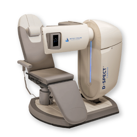


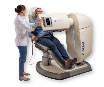
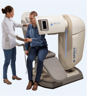
The D-SPECT open gantry design is about patient access and comfort. The scanner is designed so nuclear departments or cardiology offices can image even the most challenging patients. During a cardiac scan, such as myocardial perfusion imaging, there is no gantry motion, so patients can easily be positioned and scanned. The chair provides the flexibility to image the patient supine position, upright position or any angle in between. It supports up to 1000lb/454kg.
Spectrum Dynamics’ unique implementation of CZT solid-state detectors has established the design as the gold standard of CZT-based SPECT cameras used in Nuclear Medicine.
CZT detectors technical advantages for SPECT imaging, when compared to traditional NaI detectors, include higher sensitivity and improved energy resolution. CZT eliminates the need for photomultiplier tubes.
Spectrum Dynamics combines the CZT modules in a proprietary and patented column design. These detector columns swivel during a SPECT scan acquisition, while the gantry is in a fixed position and close to the patient There is no perceivable motion for the patient during the scanning session. This design is beneficial in more than one way: focus the field of view in a specific or an for even higher sensitivity gains during a scan acquisition; 3D dynamic imaging of any organ; lower dose protocols; scanner throughput.
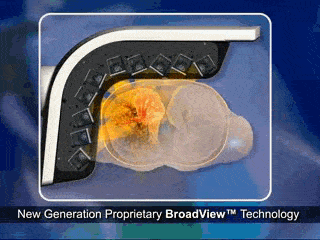
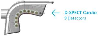
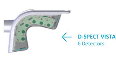
The D-SPECT camera contains 6 or 9 detector columns.
Spectrum Dynamics’ patented swivel head detectors are
pixelated to align with the parallel hole tungsten collimators
to allow for faster acquisition.
Each scanner design is optimized to allow for the gantry to be
positioned as close as possible to the patient: this ensures the
best image quality in a nuclear medicine scan: proximity with
no gantry rotation or motion.
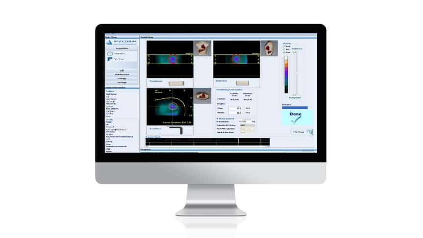
Guided Patient Positioning for Efficient Workflow
- In-gantry patient positioning guide with touch screen displays positioning information of current scan and previous scan.
- Screen displays pre-scan image for scan start confirmation.
D-SPECT: Utilizes digital technology in a unique design for high quality imaging
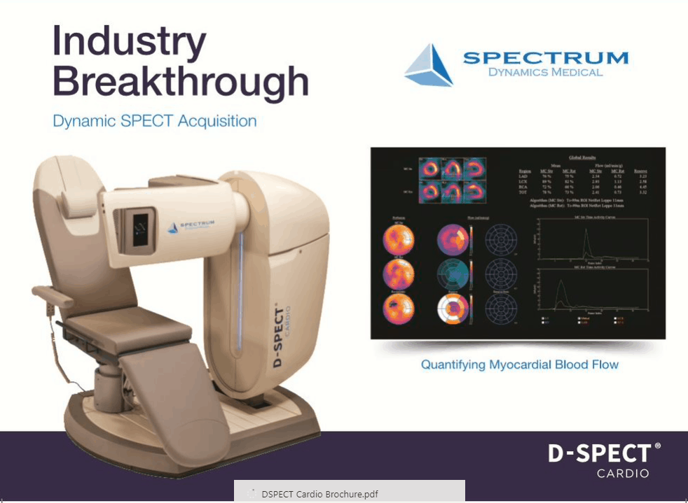
A Breakthrough in Nuclear Cardiac Imaging
Unique Approach in SPECT Myocardial Perfusion Imaging
-
List Mode Acquisition
Data can be reformatted for advanced quantification applications such CFR, motion correction, planar image -
Scout Imaging
20 sec Pre-Scan Scout based on patient’s heart -
ROI Imaging
Adaptive field of view: focus acquisition on patient’s heart vs extra cardiac activity. -
Scanning Proximity
D-SPECT design ensures proximity to the patient for high image quality, with no gantry rotation or motion
D-SPECT Image Gallery
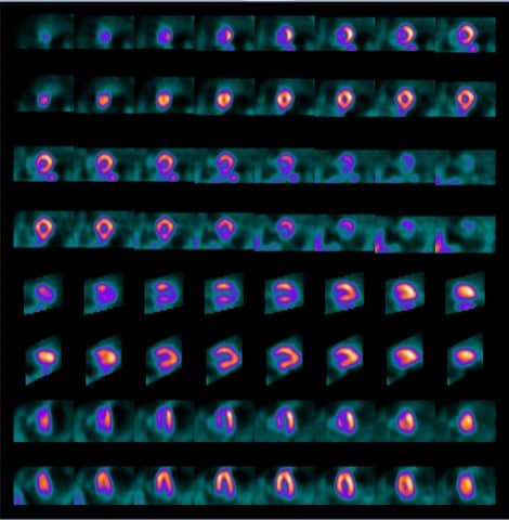
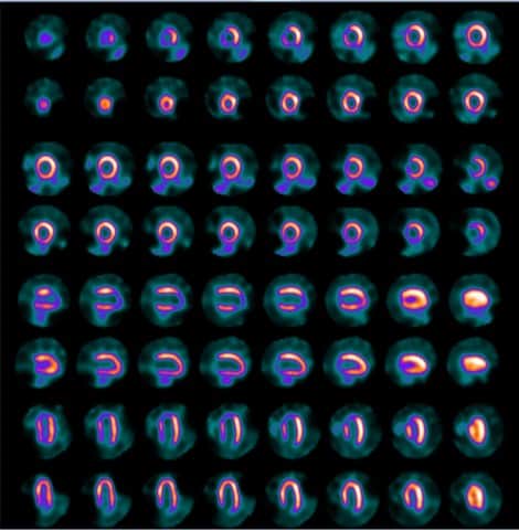
Analog
Digital

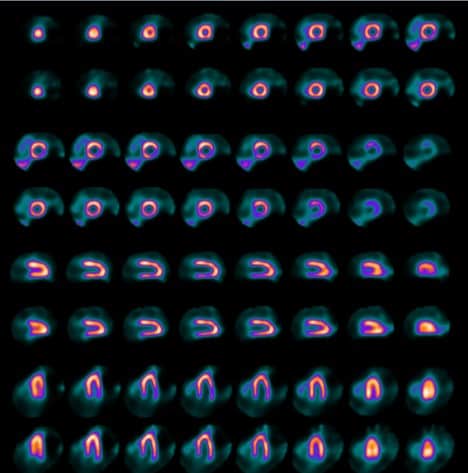
Analog
Digital
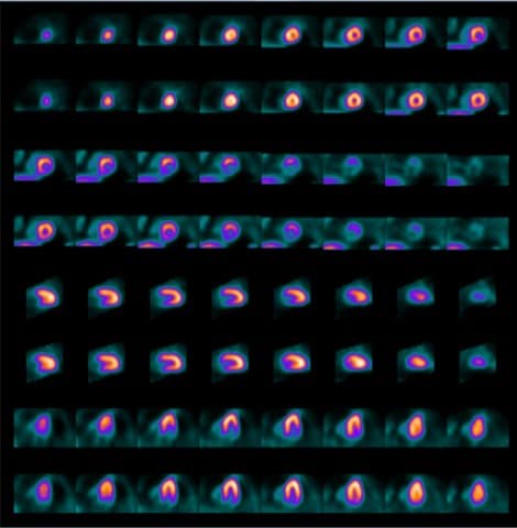
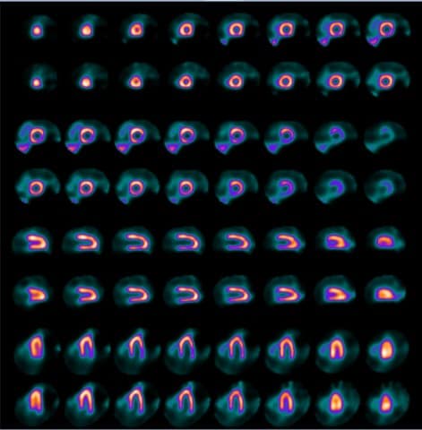
Supine
Upright
myocardial perfusion imaging
4min stress / 6min rest
myocardial perfusion imaging
4min stress / 6min rest
myocardial perfusion imaging
7min rest upright / supine
Testimonials
From the medical physicist's perspective: “The new D-SPECT CARDIO quality control is much faster and easier. The software is user-friendly, and daily QC and calibrations are very quick and easy to perform with a unique and well-defined procedure. As far as radiation protection is concerned, the D-SPECT allows injection of a lower administered activity to reduce radiation exposure while providing high-quality diagnostic images within a shorter acquisition time. It results in lower exposure to the staff.”
From the technologist's standpoint: “The D-SPECT system presents the latest generation of highly sensitive detectors arranged on a stationary gantry in a curved configuration to accommodate the patient’s heart. The unique aspect of the D-SPECT system allows patients to be imaged while sitting and positioned upright and/or supine. Upright positioning is an enhancement and an improvement for dedicated cardiac systems, especially helpful when imaging patients with chronic obstructive pulmonary disease or dyspnea, as lying flat often exacerbates shortness of breath. Superior image quality, low doses, and fast acquisition times, combined with optimal patient comfort, demonstrate that D-SPECT technology is advantageous both for the patient and the nuclear medicine professionals acquiring and reading the study."
Customer Sites
The Louis Pradel Hospital, Lyon France
Morbihan Nuclear Medicine Center (CMNM), Vannes and Lorient France
Centre Georges-François Leclerc, France
Emory University Hospital Midtown, Atlanta, USA
John Radcliffe Hospital, Oxford United Kingdom
Downloads

White Paper: Calculating Flow Reserve with CZT SPECT: Fundamentals and Applications