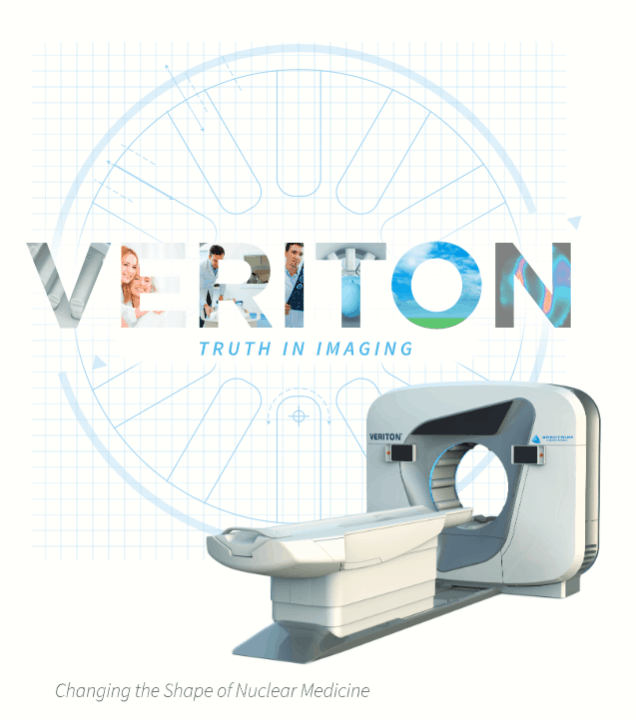
VERITON Series
First Ring-Shaped Gantry Digital SPECT/CT
360° Total Body 3D Imaging

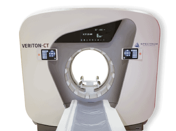
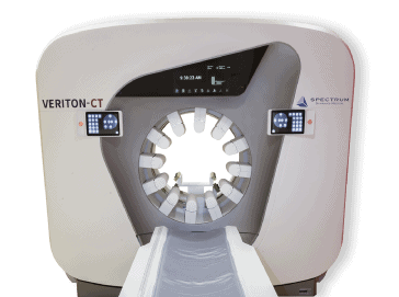
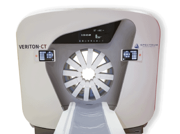
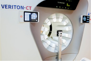
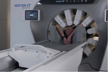
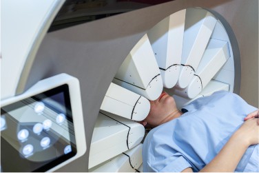
Spectrum Dynamics Medical built the VERITON with a new generation imaging technology like no other, wrapping 360° around the patient’s body contour for a personalized exam. The heart of the innovation is in a set of 12 digital CZT detectors that automatically move within millimeters of the patient’s body. The 3D hybrid digital scanner gives clinicians comprehensive information to help diagnose with confidence and accuracy.
In the past, converting a dual head camera to more expensive CZT technology without a broader clinical impact offered too little value. That’s why Spectrum Dynamics engineers took the bold step to change the shape of scanning with 360° CZT design, which improves sensitivity to a point that the system can truly affect change. Without the historical dual head design barriers, VERITON delivers proximity and efficiency, resolving complex imaging challenge without the traditional tradeoffs.
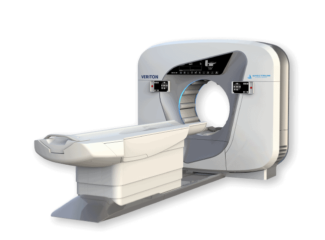
VERITON Innovative System Design: 360° CZT
Spectrum Dynamics product portfolio foundation is built on CZT solid-state digital detector technology. The goal is to maximize the benefits of CZT in routine clinical applications in nuclear medicine departments: -consistently generate clinical studies of high image quality; -shorten the scan times; -image using the lowest possible injected activity; -improve diagnostic accuracy; – A digital SPECT/CT such as the VERITON, can help drive new and innovative clinical growth in SPECT imaging with new applications only possible with digital technology, such as 3D dynamic myocardial perfusion imaging.
The engineering and algorithm teams developed four distinctive features for the optimal utilization of CZT detectors for SPECT imaging:
1. CZT columns that swivel to acquire data during acquisition
2. Unique gantry designs to optimize 3D imaging with the digital detectors as close to the organ of interest as possible, key for sensitivity and resolution gains .
3. A comprehensive reconstruction algorithm selection to generate the best image quality possible, with options that until know, were only used in PET/CT imaging for SUV calculation.
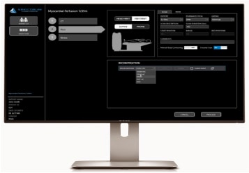
4. Integrated acquisition, quantitative reconstruction and review console that allows for workflow efficiency while enhancing the collaboration between the technologist and clinician.
First Ring-Shaped Gantry Digital SPECT/CT
Transition to Routine 3D Imaging with Next Generation SPECT imaging Technology
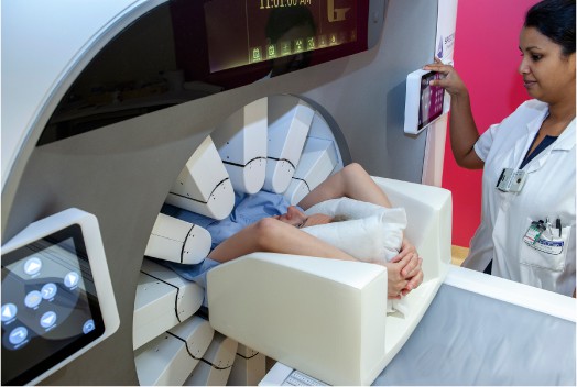
Because the digital detectors snugly surround the patient to
sense maximum photons, the 3D image quality reveals more
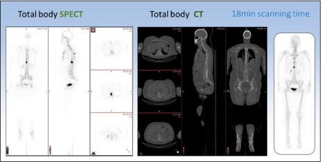
detail than planar, Anger based images. The multipurpose SPECT/CT system scans from head to toes in NM/CT hybrid mode, maximizing routine clinical utilization and providing detailed information in a single 3D scan. Avoid the need to request extra views—easier on the patient.
Total body SPECT/CT workflow, with 2m continuous coverage
during a hybrid acquisition, allows for efficiency and additional information when compared to a planar whole-body scan, plus an additional, localized SPECT/CT scan. The final 3D images are corrected for scatter and attenuation, partial volume effect and can be used to generate multi-view planar
(MVP) images, if required.
Improved lesion detection could be just the start. One total body acquisition result in a 3D view with PET-like slices,
shortening exam times, and the imaging efficiency allows a
lower injected dose. Those operational changes can transform the way imaging is scheduled and carried out.
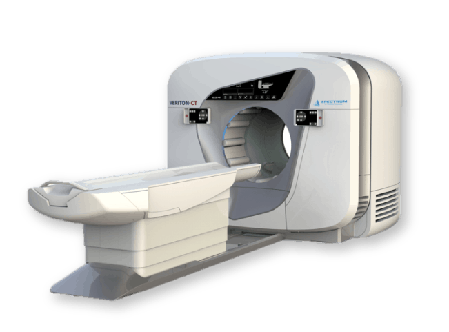
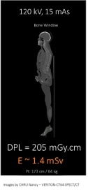
Choice of 16/64 Slice Diagnostic CT Capabilities for VERITON-CT
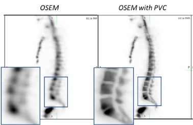
Clinicians can use the high-resolution, low-dose CT scan data, not only for attenuation correction and localization, but also to routinely reconstruct SPECT data with partial volume correction (PVC) for improved contrast and resolution. CT image quality tools, such as metal artifact reduction, LISA low-dose iterative reconstruction and 4D dose modulation, are standard features of the VERITON-CT configuration to meet the needs of demanding nuclear medicine and radiology departments.

Coverage
Up to 20/40 mm coverage with 0.625 mm spatial resolution and 2 m scan range. Clinical utility and confidence in one.

Speed
Axial and helical scans with up to 180 mmh/sec table motion and 0.5/0.4 seconds per rotation temporal sampling speed for advanced cardiac exams.

Power
Up to 500/660 mA, 5.3/8 MHU tube and 60/80 kW generator power. Adaptive range of 80 to 140 kVp for optimized protocols.
Low Dose
Improve patient care with low dose. Certified NEMA XR-29 compliant including Low Dose Iterative Reconstruction Software (LISA) and pediatric protocols.
The latest clinical innovation in digital SPECT/CT, VERITON-CT, is designed:
- For routine tomographic imaging in busy nuclear medicine departments.
- For improved diagnostic quality of nuclear medicine scans with hi-resolution CT for localization and attenuation correction.
- To accommodate patients of all sizes with its 80-cm bore size.
- For department and staff efficiency with faster 3D imaging than conventional analog camera protocol.
VERITON-CT Image Generation: A first in SPECT/CT
Console allows for true multi-tasking: acquisition, reconstruction, and quantitative post processing of the 3D data. Image tool kit includes advanced reconstruction algorithms to ensure quantitative accuracy.
The VERITON and VERITON-CT systems are equipped with
advanced image processing algorithms to ensure state-of-the-art image quality. Options include OSEM iterative reconstruction with resolution recovery, attenuation correction (AC), scatter correction (SC), partial volume correction (PVC), high energy correction (HEC) and more.
As hybrid SPECT/CT systems increasingly become the standard
of care around the globe, maximizing the use of the CT
beyond anatomical localization, attenuation correction, or
even basic diagnostic imaging becomes more relevant. Low
dose CT scan data can also be used for Partial Volume
Correction of the reconstructed image. The aim of PVC is to
transfer the sharp edges provided by the CT into the SPECT
image by constraining the activity to remain within the
anatomical region edges during the reconstruction process
Our Unique Approach to address SPECT Imaging Challenges

DESIGN
360° ring-shaped gantry design, 80cm bore
SOLID STATE TECHNOLOGY
Sensitiviy, Energy Resolution
CONFIGURATION
Swiveling, high resolution
CZT,
12 detectors
PROXIMITY
Adaptive body contouring by each detector
Image Generation
Software and Hardware Enabled Possibilities
Continued technical innovations are key to the growth of nuclear medicine as it introduces new ways to address complex imaging challenges without traditional tradeoffs. The unique approach to system geometry in VERITON, leveraging digital CZT arrays, offers improvements to photon detection and image quality. Fully digital tomographic personalized exams, enabled by swivel scanning, increase physician confidence with improved photon capture resulting in better image contrast and lesion detection. Enabled by this digital transformation of SPECT, the appeal of faster scans, lower radiation dose, fully quantitative images and improved image quality offers real potential to bring more patients back to nuclear medicine along with continued industry investment.
One Location. One Console. One Suite.
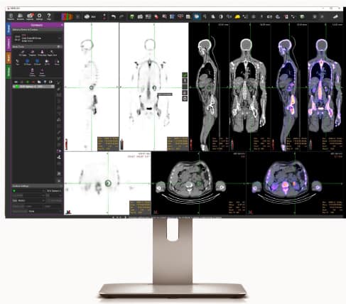
- Database Management
- Acquisition Protocol Selection
– Hybrid Protocol
– Stand-Alone Dx CT Protocol - Data QC and Reconstruction
- MIM-SD Image Review Applications
– Advanced Analysis: Quantification
– Vendor Neutral, zero-foot print solution
– Customized display workflows
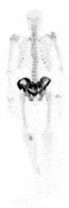
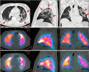
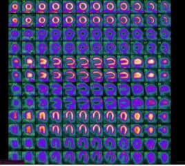
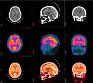
VERITON Image Gallery
Testimonials
"VERITON-CT constitutes a significant technological leap …and we are only at the beginning of exploiting its possibilities"
Customer Sites
Ridley-Tree Cancer Center in Santa Barbara, California USA
Schneider Children’s Medical Center of Israel
Institut Gustave Roussy, Paris France
King Chulalongkorn Memorial Hospital, Bangkok Thailand
University Hospital of Montreal (CHUM) – Canada
Bellvitge Hospital Universitari (HUB) – Spain
Linköping University Hospital – Sweden
Hospital Center University of Caen, France
Nancy Regional and University Hospital Center (CHRU), France
Royal United Hospitals Bath NHS Foundation Trust – United Kingdom
Downloads
White Paper: Optimized Digital Gamma Camera Design - 360° CZT for Total Body Imaging